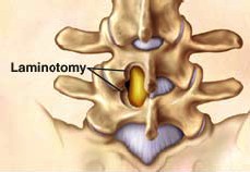
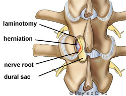
Lamina is the osseous part of the vertebra forming the ceiling of the spinal canal within which the spinal cord is located. When there is spinal canal stenosis due to spine degeneration, resulting in compression of the spinal cord or/and nerve roots exiting through intervertebral foramina, it may be necessary to remove the lamina –either totally or partially- from one side of the spine, in order to achieve decompression of the spinal canal.
In some cases, laminectomy or hemilaminectomy is essential so that the surgeon can reach the part of the intervertebral disc that is to be removed.
 Laminectomy and hemilaminectomy can be applied following modern minimally invasive surgical techniques which, unlike traditional classical methods, require much smaller incisions and are significantly less traumatic for the tissues of the operated area. The patient’s recovery and return to physical activities is very fast.
Following a wide laminectomy, instability of the spine may arise and there may be need for spine fusion at the same time in order to restore its stability.
ΤΕΧΝΙΚΗ
The surgical operation is conducted under general anaesthesia and the patient is usually discharged from the hospital the same or the next day.
 Laminectomy can be applied following three techniques:
- Mini-open. The surgeon makes as small incision through which s/he uses special instruments and a microscope.
- Tubular. During this technique, the spine is approached through an opening (and not an incision) in paraspinal muscles with a tubular dilator inserted into the skin through a very small incision. The tubular dlator serves as a “tunnel” through which special instruments pass for the application of the technique. The advantage of this method is the atraumatic approach to muscles, minimal blood loss and the impressively fast postoperative recovery and mobilization of the patient even on the very same day.
- Endoscopic. This technique is performed with the use of a micro camera called “endoscope” reaching the spine through a minimal skin inscision. The surgical field is shown on a monitor situated next to the surgical table. The surgeon performs the procedure by using special instruments while watching through the monitor and advancing them through a working cannula.
RISKS – COMPLICATIONS
As in all surgical procedures, there is the risk for infection, and there is also the risk for injury of the nerve, spinal cord or vessels of the area.
Another risk of this surgery is that, in case not enough osseous part of the lamina has been removed, there is no sufficient decompression of the spinal cord and nerve roots.
A small percentage of patients are likely to present the so-called “Failed Back Surgery Syndrome” (FBSS) with permanent postoperative spinal neuropathic pain, due to nerve root dysfunction of unknown aetiology despite the fact that the surgical procedure was performed perfectly well without any complications


