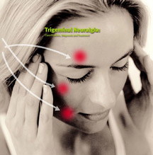
Trigeminal neuralgia (TN) is a chronic syndrome of neuropathic pain affecting the facial area. It is considered to be the most excruciating pain probably due to the fact that the facial region has a very dense distribution of sensory nerve endings per square centimeter.
The pain is so severe that it affects significantly even simple functions, such as chewing, swallowing, tooth brushing, washing, touching the face etc.
Various epidemiological studies have shown that the incidence rate of the disease is 4-5 young patients per 100.000. It is more common in the age groups between 50 and 70 years old. In 90% of cases the symptoms start at the age of 40. Women are more frequently affected compared to men in a ration 1,5:1.
The pathophysiology of trigeminal neuralgia is not clear. According to clinical observations, it is probably induced by compression of the trigeminal nerve at its origin by the brainstem, blood vessels or a tumour. The local pressure causes demyelination, which results in abnormal depolarization and finally produces ectopic pulses.
The trigeminal nerve is the fifth cranial nerve and the biggest sensory nerve covering the face and neighbouring tissues. It provides motor innervation to masseter muscles (muscles of mastication).
The nerve originates from the brainstem and enters the temporal bone where it forms the gasserian ganglion (semilunar ganglion) in a cavity of the bone which is called Meckel’s cave (trigeminal cave).
The gasserian ganglion is divided in three main branches innervating the face:
1. Opthalmic branch (first branch)
2. Maxillary branch (second branch)
3. Mandibular branch (third branch)
Trigeminal Neuralgia
DIAGNOSIS
CLINICAL DIAGNOSTIC CRITERIA
Features of pain : acute, stabbing, electric shock-like, superficial
Intensity: moderate to very severe
Duration: every attack lasts seconds. There may occur a number of different attacks almost at the same time followed by a period of calmness.
Periodicity: there may be weeks or months without pain
Localisation and distribution of pain: mainly unilaterally in the innervation region of the affected branch or branches.
Stimulating factors: light touch, talking, chewing, washing.
Relieving factors: frequent sleep, drugs
Concomitant conditions: poor quality of life, depression, weight loss
SUPPLEMENTARY EXAMINATIONS
As soon as trigeminal neuralgia is diagnosed, the patient has to undergo MRI to investigate whether there are organic pathological conditions, such as tumour or multiple sclerosis, which might cause secondary trigeminal neuralgia. Magnetic Angiography is also useful when there is suspicion for the nerve compression by a vessel. The role of vascular pressure on the nerve and the pathogenesis of neuralgia is ambiguous. In 1/3 of asymptomatic patients examined with MRI, the image revealed vascular pressure on the trigeminal nerve.
TREATMENT
A. CONSERVATIVE TREATMENT
The drug of choice is carbamazepine (Tegretol). Observation studies have demonstrated that carbamazepine (Tegretol) may reduce pain in 70% of cases. oxcarbazepine (Trileptal) slightly less effective but has fewer side effects. Alternative second line drugs, whose efficacy has not yet been established, is pregabaline (Lyrica), gabapentine (Neurontin) and baclofen (Baclofen).
PHARMACOTHERAPIES FOR TRIGEMINAL NEURALGIA
| DRUG | ΔΟΣΗ | ONSET OF RELIEF |
| CARBAMAZEPINE | 400-800 mg/day | 24-48 HOURS |
| PHENYTOINE | 300-500 mg/day | 24-48 HOURS |
| BACLOFEN | 40-80 mg/day | ? |
| CLONAZEPAM | 1,5-8 mg/day | ? |
| VALPROATE | 500-1500 mg/day | WEEKS |
| LAMOTRIGINE | 150-400 mg/day | 24 HOURS |
| PIMOZINE | 4-12mg/day | ? |
| GABAPENTIN | 900-2400 mg/day | 1 WEEK |
| OXCARBAZEPINE | 900-2800 mg/day | 24-72 HOURS |
When conservative treatment fails or drug side-effects are not tolerated, the patient has to resort to one of the invasive techniques. There are 5 clinically appropriate options:
1. Surgical Microvascular Decompression (MVD)
2. Gamma Knife Surgery (GKS)
3. Percutaneous Balloon Microcompression (PBM)
4. Percutaneous Glycerol Rhizotomy (PRGR)
5. Percutanous Radiofrequency Neurolysis of the Gasserian ganglion
6. Gasserian Ganglion Stimulation/ Neuromodulation (experimental treatment)
Surgical Microvascular Decompression (MVD). It is a surgical procedure, performed under general anaesthesia, releasing the nerve from the compressing arteries and coagulating the other microvessels involved.
Gamma Knife Treatment (GKS). It is an non-selective ablation of the gasserian ganglion by irradiation. It is applied under local anaesthesia and mild sedation. The efficacy of the method in reducing pain is limited in about 60-70%.
Percutaneous Balloon Microcompression (PBM). With this technique the trigeminal nerve is being decompressed by placing a small balloon percutaneously (through a needle) in Meckel’s cave, where the gasserian ganglion is located. This pressure causes an ischaemic injury of ganglion cells. In terms of resulting effect, the procedure is comparable with the Percutaneous Radiofrequency Neurolysis of the Gasserian ganglion. The advantage of PBM is that it is appropriate for treating neuropathy of the first trigeminal nerve branch (ophthalmic branch) keeping intact the eye corneal reflex.
Percutanous Glycerol Rhizotomy (PRGR). In this procedure, a needle is placed into the trigeminal cistern under radiological guidance. The patient is being seated with the head in flexion. For evaluating the volume of the cistern, a contrast medium is administered, which in turn is aspirated and an equal volume of glycerol is infused provoking the nerve ablation.
Percutaneous Radiofrequency Neurolysis of the Gasserian ganglion. It is preferable mainly in the elderly. The result is less satisfactory than in surgery, but this method is applied with local anaesthesia and light intravenous sedation?/ελαφριά μέθη, and has lower mortality rates.
CONCLUSION
For the young patients with clear past medical history for other concomitant diseases, the procedure of choice is surgical decompression. For the elderly who are in risk of complications from anaesthesia, the method of choice is the percutaneous radiofrequency neurolysis of the gasserian ganglion.
JOURNAL ARTICLES
Headache Classification Subcommittee of the International Headache Society. The International Classification of Heachache Disorders: 2nd edition. Cephalalgia 2004; 24 (Suppl1):9. Janneta, PJ. Microsurgical management of trigeminal neuralgia. Arch Neurol 1985; 42:800. Lance, JW. Mechanism of and management of headache, Butterworth Heinemann, Oxford 1993, p. 260. Love, S, Coakham, HB. Trigeminal neuralgia: pathology and pathogenesis. Brain 2001; 124:2347. Merskey, H, Bogduk, N. Classification of chronic pain. Descriptions of chronic pain syndromes and definitions of pain terms, IASP Press, Seattle 1994, pp. 59-71. Nurmikko, TJ, Eldridge, PR. Trigeminal neuralgia—pathophysiology, diagnosis and current treatment. Br J Anaesth 2001; 87:117. Rozen, TD, Capobianco, DJ, Dalessio, DJ. Cranial neuralgias and atypical facial pain. In: Wolff’s Headache and Other Head Pain, Siblerstein, SD, Lipton, RB, Dalessio, DJ (eds), Oxford University Press, New York 2001, pp. 509. Slavin, KV, Wess C. Trigeminal branch stimulation for intractable neuropathic pain: technical note. Neuromodulation 8:7-13, 2005
MEDICAL INFORMATION SOURCES
1. PAIN PRACTICE JOURNAL
2. BONICA”S MANAGEMENT OF PAIN
3. PAIN PHYSICIAN JOURNAL
4. INTERVENTIONAL PAIN MANAGEMENT BOOK
5. NEUROMODULATION JOURNAL


