

RECOMMENDATIONS
The treatment of shoulder pain should primarily be conservative with combination of pharmacotherapy and chiropractic/ physiotherapy. Should the above treatments fail (after being applied for a reasonable period of 4 weeks), minimally invasive techniques can be applied.

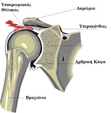
The patient is placed in the supine position. After thorough cleaning of the area with antiseptic solution, the lateral acromial angle is identified with hand palpation. A fine needle 25 gauge is inserted at the centre of this point while 4 ml of ropivacain 0,2 % & 40 mg depot medrol are injected into the bursa.
 Β. ACROMIOCLAVICULAR JOINT DYSFUNCTION
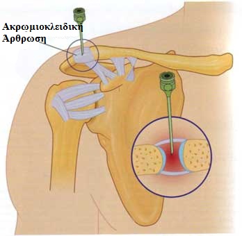
The patient is placed in the supine position. After thorough cleaning of the area with antiseptic solution, the acromioclavicular joint is identified with hand palpation 2,5cm medial to the apex of the acromion. A fine needle 25 gauge is inserted to this point while 1 ml of ropivacain 0,2% and 40 mg depot medrol are injected into the joint. While injecting, some resistance is met due to the small volume of the joint.
C. INTRA-ARTICULAR INJECTION
The patient is placed in the supine position. After thorough cleaning of the area with antiseptic solution, the middle part of the acromion is identified with hand palpation. A needle is inserted 2,5 cm below this point while 2 ml of ropivacain 0,2% and 40 mg depot medrol are injected into the joint without meeting any particular resistance.
D. PULSED RADIOFREQUENCY
APPLIED TO THE

The patient is placed in the sitting position with the head slightly bent laterally towards the opposite side. The scapular spine (spina scapulae) is hand palpated and a line is marked connecting the acromion with the medial scapular border. From the centre of this line, another line is marked parallel to the spine. The lateral superior angle is divided in half by a line. The acupuncture site is determined by the last line, in a distance of 2,5cm from the centre of the angle.
A special radiofrequency needle is inserted vertically to the skin, directed rightwards on the shoulder. In a depth of about 2,5 cm, the tip of the needle comes in contact with the scapula and the scapular notch is traced along the bone. When the needle enters the notch, an electrode is inserted through the needle.
This needle is connected with a radiofrequency generator and the suprascapular nerve is identified by conducting sensory testing. When this is localized, motor testing with low voltage electric current is performed causing contraction of the supraspinatus and infraspinatus muscles. Pulsed radiofrequency is then applied having a neuromodulatory effect on the nerve and limiting the transmission of painful signals to the shoulder. The therapy can be repeated, if necessary.
MEDICAL INFORMATION SOURCES
1. PAIN PRACTICE JOURNAL
2. BONICA”S MANAGEMENT OF PAIN
3. PAIN PHYSICIAN JOURNAL
4. INTERVENTIONAL PAIN MANAGEMENT BOOK
5. MANUAL OF RF TECHNIQUES (GAUCI)


 “Pain in Europe VII” – 7th EFIC® Congress")


