GENERAL
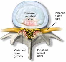
Spinal stenosis is the condition in which the spinal canal (i.e. the part of the spine enclosing the spinal cord and nerve roots) gets narrow and, as a result, exerts pressure upon the sensitive structures within the spinal canal. This condition is gradually aggravated causing pain and limiting the patient’s capacity for various physical activities. Depending on which spinal level the pressure is being exerted to the spinal cord or spinal roots, the resulting symptoms are manifested either on the neck and arms or the waist and legs.
Spinal stenosis is a quite common disorder and occurs more frequently in the elderly. It is due to the gradual, age-related, degeneration of the spine.
The spine is composed of 7 cervical, 12 thoracic, 5 lumbar, 5 fused sacral vertebrae and the coccyx. Every vertebra consists of an anterior part, the body, and a posterior part which is connected with the anterior part through the vertebral pedicle..
The posterior part includes the lamina, the spinous process (which is palpable at the back), the 2 transverse processes and the superior & inferior articular processes. The superior and inferior articular processes are interconnected bilaterally and form the facet joints, which provide further stability to the spine. The vertebrae are separated one from another with the intervertebral discs.
 Between the anterior and posterior spinal structures lies the spinal canal. Within the spinal canal are located the spinal cord and spinal roots which exit through the intervertebral foramina and course to the periphery so as to innervate the various parts of the body. When these bone structures forming the spinal canal are defected (usually due to ageing), the resulting effect is a spinal stenosis which, according to its severity, is manifested with a variety of symptoms.
SYMPTOMS
LUMBAR SPINAL STENOSIS
Low back spinal stenosis causes numbness and pain in the legs with the patient walking or/and standing up for quite a long time. The distress usually subsides when the patient bends forward or sits, and recurs when standing up and walking.
One typical example of this disorder in daily life is the patient feeling pain and leg numbness while shopping at the supermarket. When the patient leans against the trolley slightly bending forward, the symptoms are alleviated and thus s/he manages to finish the shopping.
This type of pain is entitled “neurogenic intermittent claudication” and is characteristic of spinal stenosis, as opposed to intermittent claudication of vascular aetiology which does not subside with sitting and bending forward.
Besides pain, other symptoms of low back spinal stenosis are: numbness, muscle weakness and paraesthesias (pins-and-needles) in lower limbs.
In severe cases of spinal stenosis, the patient is not able to make even one step and has urinary or defecation problems, due to intense pressure of the cauda equina, which may ultimately lead to incontinence.
CERVICAL SPINAL STENOSIS
When the stenosis is located at the cervical spine, the same symptoms as mentioned above are manifested in the neck and shoulder and may extend even down to the fingers. When the stenosis is very severe, the persisting pressure on the spinal cord causes myelopathy, that is permanent damage to the spinal cord.
INDUCTION MECHANISM OF SYMPTOMS
The normal walking increases the oxygen demand and consumption in the lower spinal nerves and leads to blood flow increase to these nerves. In healthy subjects, the oxygen supply meets the demand.
In spinal stenosis, there is impedance of the blood flow and oxygen supply to the nerves and, as a result, the oxygen supply during walking is not enough to satisfy the needs. As long as they get more and more ishaemic, the nerves start to dysfunction and this leads to pain, numbness, paraesthesia and weakness of the legs. The patient needs to sit and then the oxygen demand reduces, the supply starts becoming sufficient and when the oxygen demand coincides with the supply, the symptoms temporarily subside.
The diagnosis is set based on the very typical symptoms reported by the patient, which are confirmed with the MRI.
TREATMENT
CONSERVATIVE TREATMENT
The conservative treatment includes simple analgesics, anti-inflammatory drugs (to reduce inflammation) physiotherapy and physical exercises. In the event of radicular pain antiepileptics and antidepressants can be administered.
MINIMAL INVASIVE PAIN THERAPIES
- Transforaminal corticosteroid injection
- Caudal epidural injection
- Racz Neuroplasty
- Neuromodulation therapies
SURGICAL TREATMENT
As long as all the less invasive methods are applied without any result, there are various surgical techniques for the spinal canal decompression. These surgical techniques are conducted provided benefits outnumber potential risks and difficulties and always depending on the patient’s history case and age.
MEDICAL INFORMATION SOURCES
1. PAIN PRACTICE JOURNAL
2. BONICA”S MANAGEMENT OF PAIN
3. PAIN PHYSICIAN JOURNAL
4. INTERVENTIONAL PAIN MANAGEMENT BOOK
5. NEUROMODULATION JOURNAL


